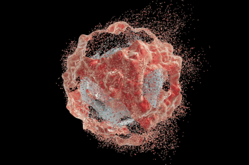
Cancer patients often face a painful trade-off: accept the harsh side effects of chemotherapy or risk letting tumors grow unchecked. The root of the problem is that most cancer drugs can’t tell healthy tissue from cancerous tissue. They spread through the body indiscriminately, attacking both good and bad cells.
A new approach from scientists at the University of Cambridge may change how this problem is tackled. Their experimental drug system remains largely inactive until two separate pieces of the drug meet each other inside tumor tissue. Only there do they snap together to form a potent compound that awakens the immune system against cancer.
The idea is simple but powerful: instead of carrying an active drug throughout the body, carry harmless fragments that only come alive where cancer is hiding.
Why the STING Pathway Is a Key Cancer Target
The treatment works by activating a cellular alarm system known as STING, short for “stimulator of interferon genes.” When triggered, STING launches waves of immune activity designed to destroy infected or damaged cells. Cancer researchers have long wanted to harness this pathway because it can turn so-called “cold” tumors, which evade immune detection, into “hot” tumors that the immune system can attack.
The challenge is that stimulating STING across the whole body can be dangerous. Earlier experimental drugs caused autoimmune reactions, where the immune system mistakenly attacked healthy tissues along with tumors. The Cambridge team set out to make a STING-targeting drug that activates only where cancer enzymes are abundant.
A Two-Part Cancer Drug That Locks Together Inside Tumors
Their solution builds on a compound called MSA2. On its own, MSA2 molecules must naturally pair up to activate STING. The researchers modified versions of MSA2 so that two different pieces, one carrying a bond-seeking group and the other a bond-accepting group, would form a permanent chemical link when they met.
These modified fragments, called N1 and E4, seek each other out and lock together much faster than typical biomedical reactions, likely because MSA2 already tends to form pairs. Once linked, they create a new compound (named SC2S) that proved about 20 times more potent than MSA2 in laboratory tests.
The clever twist is that only one piece of the drug can roam freely. The other is “caged” with a molecular cap that keeps it inactive. This cap can only be removed by an enzyme called β-glucuronidase. Tumors often produce this enzyme at higher levels than normal tissue, especially in oxygen-starved or necrotic regions. In theory, this means the drug should only activate where tumors are present.
Tests in Cells and Early Animal Models
In cultured human cells, the two-part system performed as hoped. Neither fragment alone did anything. But when β-glucuronidase was added, the cage was removed and the two fragments combined into the active drug, triggering strong immune signals even at low concentrations.
The next test was in animals. In zebrafish implanted with human breast cancer cells, the prodrug fragments on their own had little effect. Only when researchers added extra enzyme to the system did tumors shrink and cancer cells undergo programmed death. The enzyme boost also shifted immune cells called macrophages from a tumor-supporting state to a tumor-fighting state. Without the added enzyme, the zebrafish tumors didn’t produce enough β-glucuronidase on their own to activate the drug.
In mice, the story was similar. In standard mouse tumors, the treatment showed little activity, likely because enzyme levels were too low. To test the concept, the scientists engineered a mouse tumor line to overproduce β-glucuronidase. In this special model, the two-part drug successfully assembled inside tumors and accumulated there for hours while remaining nearly undetectable in healthy organs such as the liver, lungs, spleen, and kidneys.
Mice in this engineered model lived longer when given the treatment. Importantly, they also showed fewer side effects compared with a benchmark STING drug, which in this experiment caused modest decreases in red blood cells and related measures.
Limits, Challenges, and Future Cancer Drug Potential
These results, published in Nature Chemistry, are encouraging; however, there are important limitations. The zebrafish and unmodified mouse tumors did not generate enough enzyme for the prodrug to work without outside help. This means that in its current form, the system only functions reliably in tumors with very high β-glucuronidase activity. Not all cancers fall into this category.
Another practical consideration is delivery. In the mouse experiments, one drug component was injected directly into the tumor, while the other was given systemically. That works for accessible tumors, but not for cancers deep inside the body.
The authors emphasize that the true innovation is not this one drug, but the broader strategy: using two harmless fragments that only become active when combined under tumor-specific conditions. With further research, other enzymes or tumor markers could be used as triggers, potentially expanding the approach to a wider range of cancers.
If future studies confirm and extend these results, the two-part system could change how researchers think about cancer drugs. Instead of trying to design inherently “gentle” medicines, scientists might design medicines that stay inert until they reach the right environment.
This tactic could reduce side effects while maintaining or even improving effectiveness. Beyond immune-activating drugs like STING agonists, the same concept could apply to chemotherapy, targeted therapies, or other immune stimulators.
For now, though, the work remains at the proof-of-concept stage. The study involved cell cultures, zebrafish embryos, and small groups of mice — all important steps, but far from clinical use. Much larger studies, long-term safety evaluations, and eventually human trials would be needed before such a drug could ever be prescribed.
Source: https://studyfinds.org/tumor-activated-cancer-drug-shows-promise/