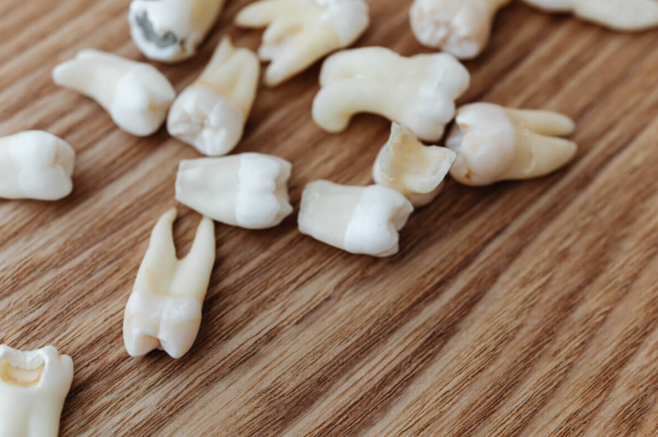
Losing several teeth changes more than just how well someone can chew a steak. A study from Japan suggests something more concerning happens. After their teeth were removed, studied animals started having memory problems. Their brains showed signs of stress and stress-related changes in the regions that handle memory and learning. What’s more, this happened even when the mice ate a normal-protein diet, suggesting the tooth loss itself, not just poor nutrition, might be affecting the brain.
Scientists at Hiroshima University tracked aging mice for six months after pulling their upper molars on both sides. They wanted to know if tooth loss leads to brain problems because people can’t eat well afterward, or if something else is going on. The answer surprised them. Mice that lost teeth performed worse on memory tests whether they ate normal or low-protein diets. When researchers examined brain tissue, they found higher levels of molecules linked to cell death, increased signs of inflammation, and fewer cells marked as neurons in key memory regions.
These are mice, not people. But the findings, published in Archives of Oral Biology, hint at an effect not explained by diet alone between missing teeth and brain changes that goes beyond just eating less protein.
How Tooth Loss Affects Memory and Cognitive Function
Researchers divided aging mice into four groups at three months old. Some had their upper molars on both sides extracted while others kept all their teeth. Half of each group received normal protein levels in their food, while the other half ate diets containing 50 percent less protein. This setup was designed to approximate what can happen for some elderly people after losing teeth: they avoid meat, fish, and eggs because chewing hurts.
Six months later, the team tested memory using a Barnes maze, a circular platform with 20 holes around its edge. Just one hole led to an escape box. Mice with intact memories learned quickly which hole offered escape. Mice with memory problems took longer to find the correct hole, often following more erratic paths.
The results were clear: mice that lost their teeth performed worse on memory tests than mice with full sets of teeth. Among mice eating less protein, this difference appeared larger, though the study notes its sample size may have been too small to detect whether diet and tooth loss interact statistically. Body weight remained steady across all groups, ruling out starvation or general poor health as explanations.
Most importantly, tooth loss drove the memory problems, whether mice ate normal-protein diets or protein-poor diets. The missing teeth themselves, not dietary protein levels, predicted which mice would struggle to remember.
Brain Cell Death Linked to Tooth Loss
The memory tests revealed problems, but brain tissue analysis showed why. Scientists examined the hippocampus, where the brain forms and stores memories. They measured gene activity for two molecules called Bax and Bcl-2 that regulate cell death pathways. When Bax gene expression rises compared to Bcl-2, it is commonly used as a marker consistent with apoptosis-related signaling, a programmed cell death process.
Mice missing teeth showed higher Bax-to-Bcl-2 ratios in their hippocampi compared to mice with intact teeth. Protein intake made no difference. Whether mice consumed normal or reduced protein, tooth loss was associated with higher levels of this apoptosis-related marker.
Additional analysis revealed markers of inflammation and fewer NeuN-positive cells in specific memory regions. The CA1 region, which helps form new memories and recall old ones, displayed high levels of GFAP and Iba-1, proteins that signal brain inflammation and stress. The same region contained fewer NeuN-positive cells, a marker consistent with reduced neuron presence in the sampled tissue.
The dentate gyrus, another memory region, showed similar patterns: markers of increased inflammation and fewer NeuN-positive cells. The CA3 region showed less change, though low protein did reduce neuron-related cell markers there. Across the hippocampal regions they examined, tooth loss had the dominant association with these brain changes while diet played a smaller role.
The Connection Between Missing Teeth and Brain Health
The relationship between oral health and brain function has interested researchers for years, though the mechanisms remain unclear.
The paper discusses several possibilities. Gum disease, which often precedes tooth loss, involves bacteria and inflammatory processes. Inflammatory signals could potentially affect blood vessels or other brain tissue. Another theory involves sensory input: teeth connect to the brain through the trigeminal nerve, one of the largest nerves in the head. Chewing sends information through this nerve to brain regions handling attention, learning, and memory. Losing teeth disrupts these signals in mice, which might affect brain activity.
This mouse study supports the idea of an effect not solely explained by nutrition. Mice fed a normal-protein diet still showed markers of brain inflammation and reduced NeuN-positive cells after tooth extraction. The tooth loss itself appears associated with brain changes in mice, not just secondary effects like malnutrition.
The research team used aging mice (SAMP8 strain) that naturally develop age-related problems including memory decline, making them useful for studying tooth loss effects in the context of aging.
Study Limitations and What They Mean
Mouse brains differ from human brains, so these findings need confirmation in people before drawing conclusions about tooth loss and dementia in humans. Mice received standardized diets while human nutrition varies widely. The six-month observation period in these aging mice may not capture all relevant time-course effects.
Reducing dietary protein meant increasing carbohydrates to maintain total calories. Higher carbohydrate intake could have influenced results, though this doesn’t change the main finding about tooth loss associations. Sample sizes ranged from seven to nine mice per group; larger studies would provide more confidence in the results, particularly regarding potential interactions between tooth loss and diet. Only male mice were tested, so whether females show the same patterns remains unknown.
Protecting Your Teeth May Protect Your Memory
In these mice, tooth loss was associated with worse memory performance, increased markers of apoptosis-related signaling, heightened neuroinflammation indicators, and fewer NeuN-positive cells in key memory regions. The tooth loss associations and the low-protein diet effects appeared to work through separate mechanisms rather than combining to amplify each other, though the authors note the study may have lacked sufficient statistical power to detect interactions confidently.
Whether similar processes occur in humans requires further study. The mouse findings suggest tooth loss could directly affect brain biology rather than working only through nutritional changes, but proving this in humans needs additional research. If the connection holds, preventing tooth loss might become one strategy for supporting cognitive health during aging.
Source : https://studyfinds.org/losing-your-teeth-fuels-cognitive-decline/